Osteoarthritis of knee

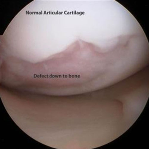
Osteoarthritis of the knee is a degenerative condition where the cartilage layer gets thinned out or eroded due to degeneration and the bone underneath it gets exposed. It is the most common cause of knee pain in the middle aged population. It is closely related to aging but not directly as a consequence of aging.
Knee joint is the largest synovial joint of the body and bears weight of the body with maximum shearing forces involved. It is formed by the lower end of thigh bone, upper end of leg bone and the kneecap, a special type of bone that forms within muscle tendon. All these three bones are covered with a thick cartilage layer, which is pain insensitive, tough, resistant to shearing force and has the capacity to absorb impact. There are two ‘C’ shaped structures placed between thigh bone and leg bone, named meniscus. Meniscus increases the congruity of the joint and also helps in absorbing impact, thus protecting the cartilage layer. The joint is filled with viscous synovial fluid which provides nutrition to the cartilage and facilitates smooth gliding of the bone surfaces during movement. Stability of the joint is maintained by different ligaments, two are inside the joint and others are on the outer surface of the joint.
Knee pain due to osteoarthritis is the second most common cause for orthopaedic outpatient visit. Eighty percent of the population above 65 years of age may have some form of osteoarthritic changes of their knee but only 60% of them need medical attention for their symptom. Females are more prone than male. There is no specific cause for osteoarthritis but some risk factors are associated with it.
- Ethnic origin
- Female gender
- Obesity
- Lifestyle and profession
- Alignment of the limb and joint
- Trauma involving the joint
- Quality of the cartilage and subchondral bone
In the beginning cartilage becomes soft and its surface starts fraying. With progress of the disease cartilage starts thinning off and ultimately erodes off exposing the underlying bone. The exposed bone rubs with each other during knee movements and weight bearing evokes pain as bone is full of pain sensitive nerve endings whereas cartilage do not have any nerve supply. This loss of cartilage height creates a lot of space between the bone surfaces which is not supported and ligament becomes lax resulting in joint instability and deformity. Initially this deformity is correctable, with progression of disease it becomes fixed.
First and common presentation is pain which is off and on in the beginning associated with stiffness on rest. Later the pain becomes persistent which aggravates with activity and subsides on rest. There may be swelling of the joint. As the disease progresses the joint starts getting deformed and movement restricted. In the very advance stage it may become awkwardly deformed for mobility.
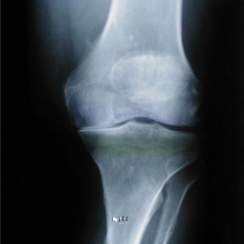
X-ray of the knee in three planes to evaluate the joint space in stress is the primary requirement. MRI may require an early stage when x-ray is not conclusive or symptoms not matched with x-ray presentation. MRI can detect meniscal injury or torn cartilage flap which can be the cause of acute pain. Blood tests are done to rule out any inflammatory condition or other cause of arthritis. X-ray wise osteoarthritis is described in 4 grades 0 to 4. Grade 0 is normal knee whereas Garde 4 is advanced stage with total loss of cartilage. Grade 4 is very wide in representation starting from focal loss of cartilage reasonably shaped knee to totally deformed mangled knee.
In early OA treatment is analgesic, ice pack, exercise and lifestyle modification. surgical treatment may be required to prevent the progression of disease and improve symptoms. These surgeries include meniscal repair and osteotomy to realign the weight bearing. In full thickness cartilage degeneration, the treatment of choice is joint replacement either total or partial.


