Knee Osteotomy

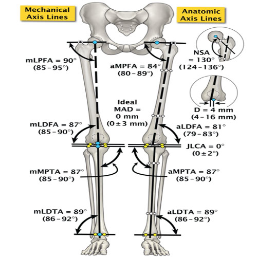
Axis line of both lower limb to determine the deformity and alignment of lower limb
For which conditions osteotomy around the knee joint is done and How?
For different conditions hip osteotomy is done. The most common conditions are:
- Correction of deformity, like
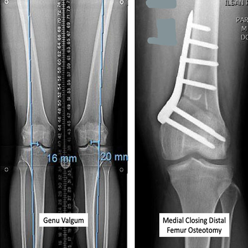
i. Genu Valgum: developmental, post rickets sequel
ii. Genu Varum: developmental, post rickets sequel
iii. Genu Recurvetum
iv. Post-traumatic malunion: there is no specific pattern of deformity and may need multiplane correction.
To correct the deformity the first step is to find out from where the deformity is originating. It can be around the joint from the metaphyseal area or intra-articular or from diaphysis. Investigation of choice is ortho scanogram, where a full length x-ray covering hip to ankle in standing position is done and then alignments are checked by drawing different angles between long axis of bone and joint surface. Based on the ortho scanogram measurement source of deformity is determined as well as amount of deformity also. Next step is surgical planning, osteotomy is planned for the bone which is contributing to the deformity of either distal femur or proximal tibia or the both. If the deformity is coming from one bone and osteotomy is performed in the other bone then limb deformity will correct but the joint alignment will get changed and will put it under undue stress.
After determining the site of deformity, the site of osteotomy is planned and the amount of bone resection for closing wedge and amount of angular distraction in case of opening wedge osteotomy is calculated to perform during surgery. Based on this pre operative calculation osteotomy is being done and fixed with anatomical plate specified for that osteotomy fixation. Depending on the intra operative stability and bone strength, the patient is allowed to bear weight and mobilise. After 6-8 weeks of surgery when the osteotomy gets united and evidenced on x-ray. full weight bearing without any walking aid is being allowed. For distal femur closing wedge osteotomy and for proximal tibia opening wedge osteotomy is being performed most commonly
b. Prevention of knee osteoarthritis and symptomatic relief:
In early medial compartment osteoarthritis of knee long axis of weight transfer get shifted more medially due to collapse of medial joint and development of varus deformity. This phenomenon contributes to more weight transfer on the diseased surface of the joint. This causes more symptoms like pain and also enhances the progression of arthritis as the joint surface gets more damaged due to excessive weight transfer. In this situation if the weight bearing axis is shifted laterally the diseased medial condyle get offloaded and pain get relieved as less weight is being transferred.
c. Provide stability of the joint by changing the slope of tibia when ligament reconstruction is not possible.
d. Prevent graft failure after ligament reconstruction by changing tibial slope and varus angulation.

